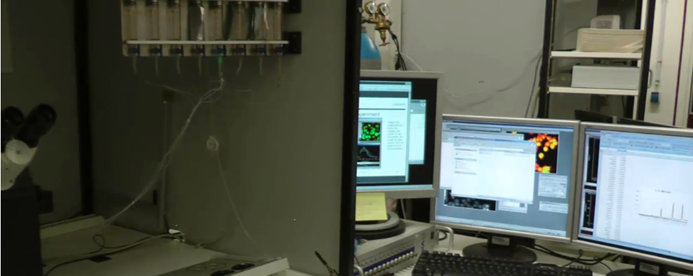
Creative Bioarray
  • USA:
  • Europe:
LOGO
  • Home
    • About Us
    • Contact Us
    • Events
  • Screening
    • Ionic Screening Service
      • Ion Flux Assay
      • Fluorescence Assay
      • Patch-clamp Assay
    • Ionic Screening Panel
      • Sodium Channels
      • Potassium Channels
      • Chloride Channels
      • Calcium Channels
      • TRP Channels
      • ATP gated P2X Channels
      • ASICs
      • Nicotinic Acetylcholine Receptors
      • Ionotropic Glutamate-gated Receptors
      • GABAa Receptors
      • Glycine Receptors
      • 5-HT Receptors3
      • Cystic Fibrosis Transmembrane Conductance Regulators (CFTR)
      • Other Ion Channels
    • Stable Cell Lines
      • Ion Channel Expressing Cell
      • GPCR Expressing Cell
      • Other Membrane Receptors/Transporters/Exchangers Expression Cell
      • Customized Ionic Channel Expression System
  • Cardiology
    • Cardiac Ion Channel Screen Panels
      • HERG (IKR, KV11.1)
      • Cav1.2
      • Kv4.3
      • KCNQ1/mink/Kv7.1
      • Kir2.1
      • Nav1.5
      • Kv1.5
      • HCN1&HCN4
      • Other Ion Channels
    • CiPA
      • CiPA Cardiac Channel Assays
      • CiPA In silico Modeling
      • CiPA Translational Assays
    • 3D Cardiotoxicity
    • Cardiomyocyte Functions
      • Action Potential Properties
      • Calcium Transient in Cardiomyocytes
      • QT Service Using Langendorff Perfused Heart
    • Cardiac in vivo Assays
      • QRS, QT Waves Detection by ECG
  • Neurology
    • Neurotransmission Functions
      • sEPSC/sIPSC Recordings
      • fEPSP / Populational Spikes Recordings
      • LTP & LTD Formation or Deficit Detection
    • Basic Neuronal Activity
      • Action Potential Properties Assay
      • NMDA/AMPA Ratio Test
      • Calcium Signaling Test
      • Customized Electrophysiological Test
    • Neuronal in vivo Assays
      • In vivo Extra-cellular Recordings/Whole-cell Patch-clamp Recordings
      • Continuous Telemetric EEG Recordings
    • Animal Behavior Tests
      • Spontaneous Locomotor Functions
      • Cognitive Functions
      • Anxiety-Depression and Social Interaction Evaluation
      • Assays for Neuropsychiatric Disorders
  • Ophthalmology
  • Platform
    • Experiment Systems
      • Xenopus Oocyte Screening Model
      • Acute Isolated Cardiomyocytes
      • Acute Dissociated Neurons
      • Primary Cultured Neurons
      • Cultured Neuronal Cell Lines
      • iPSC-derived Cardiomyocytes/Neurons
      • Acute/Cultured Organotypic Brain Slices
      • Oxygen Glucose Deprivation Model
      • 3D Cell Culture
      • iPSC-derived Neurons
      • Isolation and culture of neural stem/progenitor cells
    • Animal Models
      • Alzheimer's Disease Model
      • Parkinson's Disease Model
      • Huntington's Disease Model
      • Epilepsy Model
      • ALS Model
      • Psychiatric Model
      • Autism Spectrum Disorder
      • Cerebral-Spinal Injury Model
      • Pain Model
      • Stroke Model
      • Fragile X Model
      • Acute/Chronic Heart Failure Model
    • Techinques
      • Manual Patch-clamp Technique
      • Automated Patch-clamp
      • Multi-Electrode Array (MEA)
      • FluxOR™ Thallium Assay
      • FLIPR Detection System
      • Optogenetics
      • Neuronal Tract Tracing
    • Resource
      • Technical Methods
      • Neuroscience
      • Channelopathies
      • Channelomics
    • Equipment
  • Order
  • Careers
  • Home
  • Platform
  • Equipment
  • Screening
  • Ionic Screening Service Ionic Screening Panel Stable Cell Lines
  • Cardiology
  • CiPA 3D Cardiotoxicity Cardiomyocyte Functions Cardiac in vivo Assays Cardiac Ion Channel Screen Panels
  • Neurology
  • Basic Neuronal Activity Neurotransmission Functions Neuronal in vivo Assays Animal Behavior Tests
  • Platform
  • Experiment Systems Animal Models Techinques Resource Equipment

Equipment

The AcronTM instrument portfolio provides a comprehensive solution for patch-clamping that includes amplifiers, digitizer, software, and accessories. Our advanced instruments facilitate the entire range of patch-clamp electrophysiology techniques from the smallest single-channel to the largest macroscopic recordings.

Equipment

Patch Clamp Amplifier

Patch-clamp electrophysiology is performed with a borosilicate electrode of 1-2 µm tip diameter within a pipette containing electrolyte solution. The pipette tip is tightly sealed onto the membrane and "patches" an isolated part of the membrane, electrically. Currents flow through the channels in this patch and can then be recorded by an electrode connected to a differential amplifier. The amplifier contains the circuitry necessary to measure current passing through the cell membrane both in magnitude and direction. The pipette tip makes a giga-ohm seal with the membrane, providing metrics related to membrane conductance from the electrical current flow.

Digitizer

The current acquired by the amplifier is an analog signal, but in order to perform data analysis needed for high resolution patch-clamp measurements, the analog signal must be converted into a digital one. The digitizer is a data acquisition instrument that converts analog signals into digital signals.

Software

Analysis software is an interface with the amplifier, digitizer, and any other patch-clamp electronics, to perform data acquisition and data analysis, as well as to control the digitizer and amplifier.

Headstage

The electrical signal acquired by the micropipette needs to be transmitted to amplifier systems for signal processing. Each headstage is specifically tuned for the amplifier. A device that holds the micropipettes with built-in circuitry to transmit electrical signals from the micropipettes onto the amplifier.

Microscope

The microscope is an important optical magnification tool. The micromanipulator is a device that mechanically maneuvers the micropipette with nanometer precision, typically allowing 3D movements. The microscope with micromanipulator is to precisely and stably position the micropipette to the area of the cell membrane. It's required for a successful recording. An inverted microscope is preferable because it allows easier access for electrodes from above the preparation and also provides a larger, more solid platform to bolt the micromanipulator.

Faraday Cage and Tables

A Faraday cage is a wire mesh enclosure around your microscope and recording chamber to prevent the electrodes from picking up extraneous noise sources. The air or anti-vibration tables are used to isolate setup from external sources of vibration that may disrupt this alignment.

Creative Bioarray offers a comprehensive portfolio of electrophysiology studies to monitor neuron-to-neuron communication and study the effects of physiological or pharmacological stimuli. Small electrical signals of action, membrane, or synaptic potentials are amplified and recorded using state-of-the-art equipment.


For research use only.

Related Section

  • Experiment Systems
  • Animal Models
  • Techinques
  • Resource

Inquiry


Subscribe
  • Links
  • Sodium Channels
  • Calcium Channels
  • Potassium Channels
  • Chloride Channels
  • TRP Channels
  • ATP gated P2X Channels
  • ASICs
  • USA
  • Europe

Copyright © 2025 Creative Bioarray. All rights reserved.