Creative Bioarray
  • USA:
  • Europe:
LOGO
  • Home
    • About Us
    • Contact Us
    • Events
  • Screening
    • Ionic Screening Service
      • Ion Flux Assay
      • Fluorescence Assay
      • Patch-clamp Assay
    • Ionic Screening Panel
      • Sodium Channels
      • Potassium Channels
      • Chloride Channels
      • Calcium Channels
      • TRP Channels
      • ATP gated P2X Channels
      • ASICs
      • Nicotinic Acetylcholine Receptors
      • Ionotropic Glutamate-gated Receptors
      • GABAa Receptors
      • Glycine Receptors
      • 5-HT Receptors3
      • Cystic Fibrosis Transmembrane Conductance Regulators (CFTR)
      • Other Ion Channels
    • Stable Cell Lines
      • Ion Channel Expressing Cell
      • GPCR Expressing Cell
      • Other Membrane Receptors/Transporters/Exchangers Expression Cell
      • Customized Ionic Channel Expression System
  • Cardiology
    • Cardiac Ion Channel Screen Panels
      • HERG (IKR, KV11.1)
      • Cav1.2
      • Kv4.3
      • KCNQ1/mink/Kv7.1
      • Kir2.1
      • Nav1.5
      • Kv1.5
      • HCN1&HCN4
      • Other Ion Channels
    • CiPA
      • CiPA Cardiac Channel Assays
      • CiPA In silico Modeling
      • CiPA Translational Assays
    • 3D Cardiotoxicity
    • Cardiomyocyte Functions
      • Action Potential Properties
      • Calcium Transient in Cardiomyocytes
      • QT Service Using Langendorff Perfused Heart
    • Cardiac in vivo Assays
      • QRS, QT Waves Detection by ECG
  • Neurology
    • Neurotransmission Functions
      • sEPSC/sIPSC Recordings
      • fEPSP / Populational Spikes Recordings
      • LTP & LTD Formation or Deficit Detection
    • Basic Neuronal Activity
      • Action Potential Properties Assay
      • NMDA/AMPA Ratio Test
      • Calcium Signaling Test
      • Customized Electrophysiological Test
    • Neuronal in vivo Assays
      • In vivo Extra-cellular Recordings/Whole-cell Patch-clamp Recordings
      • Continuous Telemetric EEG Recordings
    • Animal Behavior Tests
      • Spontaneous Locomotor Functions
      • Cognitive Functions
      • Anxiety-Depression and Social Interaction Evaluation
      • Assays for Neuropsychiatric Disorders
  • Ophthalmology
  • Platform
    • Experiment Systems
      • Xenopus Oocyte Screening Model
      • Acute Isolated Cardiomyocytes
      • Acute Dissociated Neurons
      • Primary Cultured Neurons
      • Cultured Neuronal Cell Lines
      • iPSC-derived Cardiomyocytes/Neurons
      • Acute/Cultured Organotypic Brain Slices
      • Oxygen Glucose Deprivation Model
      • 3D Cell Culture
      • iPSC-derived Neurons
      • Isolation and culture of neural stem/progenitor cells
    • Animal Models
      • Alzheimer's Disease Model
      • Parkinson's Disease Model
      • Huntington's Disease Model
      • Epilepsy Model
      • ALS Model
      • Psychiatric Model
      • Autism Spectrum Disorder
      • Cerebral-Spinal Injury Model
      • Pain Model
      • Stroke Model
      • Fragile X Model
      • Acute/Chronic Heart Failure Model
    • Techinques
      • Manual Patch-clamp Technique
      • Automated Patch-clamp
      • Multi-Electrode Array (MEA)
      • FluxOR™ Thallium Assay
      • FLIPR Detection System
      • Optogenetics
      • Neuronal Tract Tracing
    • Resource
      • Technical Methods
      • Neuroscience
      • Channelopathies
      • Channelomics
    • Equipment
  • Order
  • Careers
  • Home
  • Platform
  • Resource
  • Technical Methods
  • Screening
  • Ionic Screening Service Ionic Screening Panel Stable Cell Lines
  • Cardiology
  • CiPA 3D Cardiotoxicity Cardiomyocyte Functions Cardiac in vivo Assays Cardiac Ion Channel Screen Panels
  • Neurology
  • Basic Neuronal Activity Neurotransmission Functions Neuronal in vivo Assays Animal Behavior Tests
  • Platform
  • Experiment Systems Animal Models Techinques Resource Equipment

Patch-Clamp Recording Protocol

The major limitation of in vivo patch-clamp recording is its technical difficulty, which requires experienced personnel and a large amount of patience. The workflow of patch-clamp recording involves pressing a glass micropipette against a cell in order to isolate a small "patch" of membrane that contains one or more ion channels. The experimental setup further allows researchers to "clamp" the electrical environment of the patched area by precisely controlling the voltage across the cell membrane, which, depending on the ion channels present, impacts the flow of ions through the membrane and allow for intricate study of these channels.

Patch-Clamp Recording Protocol

Here we go over the steps required to perform a patch clamp recording.

Start by pulling a borosilicate glass tube into micropipettes using a pipette puller.

Next, fire polish the tip to obtain the appropriate diameter and resistance.

After polishing, fill the micropipette with an ionic solution and flick gently to dislodge any air bubbles.

Then slide the micropipette over the electrode attached to a holder.

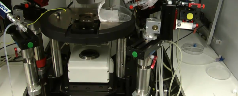
Once attached, use a syringe to apply positive pressure to the pipette, which prevents other solutions from entering the tip.

Place your cells or tissue of interest on the microscope stage and move the micropipette towards a cell.

With the amplifier generating test voltage pulses, record the resistance, which will increase once the tip is touching the cell.

To form the gigaohm seal, gently switch from positive to negative pressure using the syringe. The formation of the seal will result in a rapid increase in resistance to greater than 1 gigaohm.

Now that a cell-attached configuration has been established, and then convert to a whole-cell configuration when the membrane is ruptured. The technique is particularly applicable to small cells in the size range of 5‐20 µm in diameter, and yields good recordings in cells as small as red blood cells.

Rupturing is accomplished by adding negative pressure to the micropipette. To be more specific, negative pressure is applied to form a giga seal. Suction or a "zap" voltage pulse can be applied to break the membrane. A set of programmable valves control these pressure-switch operations.

Once the membrane breaks, the test pulse shape will have large current transients, as the cell membrane is now acting as a capacitor, which is charged by the test pulse.

The properties of a single ion channel type in any given neuron can be investigated by blocking the activity of other channels types pharmacologically.

A voltage-step protocol is used to examine ion channel currents evoked by stepping the voltage to a series of different holding potentials.

The current-voltage or IV curve shows the voltage-dependences of current flowing through an ion channel and provides insight into at which voltages the channel is open or closed.

Key Factors of a Successful Patch-Clamp Experiment

  • Clean pipette tip
  • The formation of a giga seal
  • Proper positive pressure during penetration
  • Successful membrane break-in

Expectations of Patch-Clamp Recording

It has been attracting more attention in the neuroscience field with electrophysiological techniques. Particularly for researchers who are interested in intact neural circuits, in vivo patch clamp recording could be very helpful if properly combined with other tools, such as optogenetics and two-photon imaging.

References

  1. Patch Clamp Electrophysiology, JoVE Science Education Library.
  2. Can Tao, et al. Functional dissection of synaptic circuits: in vivo patch-clamp recording in neuroscience. Front. Neural Circuits. 2015.
For research use only.

Related Section

  • Neuroscience
  • Channelopathies
  • Channelomics

Inquiry


Subscribe
  • Links
  • Sodium Channels
  • Calcium Channels
  • Potassium Channels
  • Chloride Channels
  • TRP Channels
  • ATP gated P2X Channels
  • ASICs
  • USA
  • Europe

Copyright © 2025 Creative Bioarray. All rights reserved.