Creative Bioarray
  • USA:
  • Europe:
LOGO
  • Home
    • About Us
    • Contact Us
    • Events
  • Screening
    • Ionic Screening Service
      • Ion Flux Assay
      • Fluorescence Assay
      • Patch-clamp Assay
    • Ionic Screening Panel
      • Sodium Channels
      • Potassium Channels
      • Chloride Channels
      • Calcium Channels
      • TRP Channels
      • ATP gated P2X Channels
      • ASICs
      • Nicotinic Acetylcholine Receptors
      • Ionotropic Glutamate-gated Receptors
      • GABAa Receptors
      • Glycine Receptors
      • 5-HT Receptors3
      • Cystic Fibrosis Transmembrane Conductance Regulators (CFTR)
      • Other Ion Channels
    • Stable Cell Lines
      • Ion Channel Expressing Cell
      • GPCR Expressing Cell
      • Other Membrane Receptors/Transporters/Exchangers Expression Cell
      • Customized Ionic Channel Expression System
  • Cardiology
    • Cardiac Ion Channel Screen Panels
      • HERG (IKR, KV11.1)
      • Cav1.2
      • Kv4.3
      • KCNQ1/mink/Kv7.1
      • Kir2.1
      • Nav1.5
      • Kv1.5
      • HCN1&HCN4
      • Other Ion Channels
    • CiPA
      • CiPA Cardiac Channel Assays
      • CiPA In silico Modeling
      • CiPA Translational Assays
    • 3D Cardiotoxicity
    • Cardiomyocyte Functions
      • Action Potential Properties
      • Calcium Transient in Cardiomyocytes
      • QT Service Using Langendorff Perfused Heart
    • Cardiac in vivo Assays
      • QRS, QT Waves Detection by ECG
  • Neurology
    • Neurotransmission Functions
      • sEPSC/sIPSC Recordings
      • fEPSP / Populational Spikes Recordings
      • LTP & LTD Formation or Deficit Detection
    • Basic Neuronal Activity
      • Action Potential Properties Assay
      • NMDA/AMPA Ratio Test
      • Calcium Signaling Test
      • Customized Electrophysiological Test
    • Neuronal in vivo Assays
      • In vivo Extra-cellular Recordings/Whole-cell Patch-clamp Recordings
      • Continuous Telemetric EEG Recordings
    • Animal Behavior Tests
      • Spontaneous Locomotor Functions
      • Cognitive Functions
      • Anxiety-Depression and Social Interaction Evaluation
      • Assays for Neuropsychiatric Disorders
  • Ophthalmology
  • Platform
    • Experiment Systems
      • Xenopus Oocyte Screening Model
      • Acute Isolated Cardiomyocytes
      • Acute Dissociated Neurons
      • Primary Cultured Neurons
      • Cultured Neuronal Cell Lines
      • iPSC-derived Cardiomyocytes/Neurons
      • Acute/Cultured Organotypic Brain Slices
      • Oxygen Glucose Deprivation Model
      • 3D Cell Culture
      • iPSC-derived Neurons
      • Isolation and culture of neural stem/progenitor cells
    • Animal Models
      • Alzheimer's Disease Model
      • Parkinson's Disease Model
      • Huntington's Disease Model
      • Epilepsy Model
      • ALS Model
      • Psychiatric Model
      • Autism Spectrum Disorder
      • Cerebral-Spinal Injury Model
      • Pain Model
      • Stroke Model
      • Fragile X Model
      • Acute/Chronic Heart Failure Model
    • Techinques
      • Manual Patch-clamp Technique
      • Automated Patch-clamp
      • Multi-Electrode Array (MEA)
      • FluxOR™ Thallium Assay
      • FLIPR Detection System
      • Optogenetics
      • Neuronal Tract Tracing
    • Resource
      • Technical Methods
      • Neuroscience
      • Channelopathies
      • Channelomics
    • Equipment
  • Order
  • Careers
  • Home
  • Platform
  • Resource
  • Channelopathies
  • Screening
  • Ionic Screening Service Ionic Screening Panel Stable Cell Lines
  • Cardiology
  • CiPA 3D Cardiotoxicity Cardiomyocyte Functions Cardiac in vivo Assays Cardiac Ion Channel Screen Panels
  • Neurology
  • Basic Neuronal Activity Neurotransmission Functions Neuronal in vivo Assays Animal Behavior Tests
  • Platform
  • Experiment Systems Animal Models Techinques Resource Equipment

Potassium-aggravated Myotonia

Potassium aggravated myotonia is a disease that affects muscles used for exercise. The disease usually starts in childhood or adolescence. People with this disease will continue to show a continuous burst of muscle tension (muscle stiffness) that prevents the muscles from relaxing normally. This can cause muscle stiffness, and it can increase muscle stiffness after exercise and eating potassium-rich foods (such as bananas and potatoes). This phenomenon occurs in the skeletal muscles of the whole body. The severity of potassium-aggravated myotonia ranges from mild muscle stiffness attacks to severe disabling diseases (frequent attacks). Unlike some other forms of myotonia, potassium-enhanced myotonia usually has no symptoms of muscle weakness.

Potassium-aggravated Myotonia

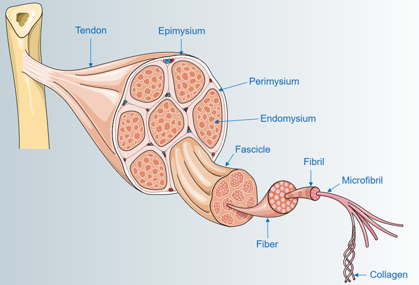
Figure 1. Muscle structure diagram.

Pathogenesis

Through clinical research and analysis, potassium aggravated myotonia is caused by mutations in the SCN4A gene. The SCN4A gene belongs to a gene family that encodes the constituent proteins of sodium channels. It is well known that in order for the body to move normally, skeletal muscles must be tense (contracted) and relaxed in a coordinated manner. Muscle contraction is triggered by the influx of positively charged atoms (ions) including sodium into skeletal muscle cells. The SCN4A protein is an important part of these cell channels that control the influx of sodium ions. These channels transport positively charged sodium atoms (sodium ions) into the cell, and play a key role in the cell's ability to generate and transmit electrical signals. The SCN4A gene provides instructions for the production of key parts (subunits) of sodium channels, which are abundant in muscles (skeletal muscles) used for exercise.

Mutations in the SCN4A gene change the general structure and function of sodium channels. The changed channel cannot properly adjust the ion flow, thereby increasing the movement of sodium ions into the skeletal muscle cells. The influx of excess sodium ions causes muscle contraction, which is a sign of muscle rigidity. Several mutations in the SCN4A gene can cause potassium to aggravate myotonia, which can cause myotonia attacks and prevent normal muscle relaxation. Eating potassium-rich foods may increase muscle stiffness. The most common genetic change associated with potassium aggravated myotonia is the substitution of the amino acid glycine for one of several other amino acids at position 1306 of the protein. These mutations delay the closure of the channel formed by the SCN4A protein, thereby increasing the flow of sodium ions into skeletal muscle cells. The presence of excess potassium will stimulate more sodium ions to flow into these cells. These changes in ion migration trigger prolonged muscle contraction, which is the basis for the muscle stiffness characteristic of potassium-aggravated myotonia.

In addition, myotonia increased by potassium ions is genetically manifested as an autosomal dominant inheritance. The patient presents with severe and lasting muscle stiffness or fluctuating muscle stiffness, which is more common within 20 minutes after exercise. Eating potassium-rich food can induce muscle stiffness, and the disease is not sensitive to cold. Clinically, there are a large class of diseases similar to the clinical symptoms of myotonia aggravated by potassium ion, and there are mutations in the SCN4A gene, mainly including fluctuating myotonia (G1y1306A1a and Ser804Phe), persistent myotonia (Gly130Glu), and acetazolamide-responsive myotonia (I1e1160Va1), and painful congenital myotonia (Va1445Met), etc.

References

  1. Colding-Jørgensen E, et al. Autosomal dominant monosymptomatic myotonia permanens. Neurology. 2006, 67(1):153-5.
  2. Lerche H, et al. Human sodium channel myotonia: slowed channel inactivation due to substitutions for a glycine within the III-IV linker. J Physiol. 1993, 470:13-22.
  3. Vicart S, et al. Human skeletal muscle sodium channelopathies. Neurol Sci. 2005, 26(4):194-202.
For research use only.

Related Section

  • Technical Methods
  • Neuroscience
  • Channelomics

Inquiry


Subscribe
  • Links
  • Sodium Channels
  • Calcium Channels
  • Potassium Channels
  • Chloride Channels
  • TRP Channels
  • ATP gated P2X Channels
  • ASICs
  • USA
  • Europe

Copyright © 2025 Creative Bioarray. All rights reserved.