- Home
-
Screening
- Ionic Screening Service
-
Ionic Screening Panel
- Ligand Gated Ion Channels
- Glycine Receptors
- 5-HT Receptors3
- Nicotinic Acetylcholine Receptors
- Ionotropic Glutamate-gated Receptors
- GABAa Receptors
- Cystic Fibrosis Transmembrane Conductance Regulators (CFTR)
- ATP gated P2X Channels
- Voltage-Gated Ion Channels
- Calcium Channels
- Chloride Channels
- Potassium Channels
- Sodium Channels
- ASICs
- TRP Channels
- Other Ion Channels
- Stable Cell Lines
- Cardiology
- Neurology
- Ophthalmology
-
Platform
-
Experiment Systems
- Xenopus Oocyte Screening Model
- Acute Isolated Cardiomyocytes
- Acute Dissociated Neurons
- Primary Cultured Neurons
- Cultured Neuronal Cell Lines
- iPSC-derived Cardiomyocytes/Neurons
- Acute/Cultured Organotypic Brain Slices
- Oxygen Glucose Deprivation Model
- 3D Cell Culture
- iPSC-derived Neurons
- Isolation and culture of neural stem/progenitor cells
- Animal Models
- Techinques
- Resource
- Equipment
-
Experiment Systems
- Order
- Careers
Romano-Ward Syndrome
Romano-Ward syndrome is a disease that causes the normal rhythm of the heart (arrhythmia) to be interrupted. This disease is a form of prolonged QT syndrome, which is a type of heart disease that causes the heart (heart) muscles to spend more time than usual to replenish the heartbeat. Among them, "long QT" refers to the specific pattern of heart activity detected by electrocardiogram (ECG or EKG). In patients with long QT syndrome, the abnormally prolonged part of the heartbeat in the QT interval leads to abnormal heart recovery time, which in turn causes abnormal heart rhythm.
Arrhythmias associated with Romano-Ward syndrome can cause syncope (syncope) or cardiac arrest and sudden death. However, there are some people with Romano-Ward syndrome who have never encountered health problems related to the disease. According to its genetic causes, 15 long QT syndromes have been identified. Certain types of long QT syndrome involve other heart abnormalities or other body system problems. Romano-Ward syndrome includes those types that involve only a longer QT interval and no other abnormalities.

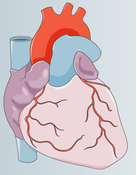
Figure 1. Heart structure diagram.
Pathogenesis
In Romano-Ward syndrome, mutations in the KCNQ1, KCNH2, and SCN5A genes are the most common causes.
KCNQ1
KCNQ1 gene belongs to a large family that provides genes for preparing potassium ion channel instructions. The KCNQ1 protein interacts with proteins in the KCNE family (such as KCNE1 protein) to form a functional potassium channel. Among them, the four alpha subunits made of KCNQ1 protein form the structure of each channel. KCNE protein directs the synthesis of β subunits,which binds (combine) with the channel to regulate its activity. These channels transport the positively charged atoms (ions) of potassium out of the cell, and play a key role in the cell's ability to generate and transmit electrical signals.
The specific function of potassium channel depends on its protein composition and its location in the body. The channels involved in KCNQ1 protein mainly exist in the inner ear and heart (heart) muscle. In the inner ear, these channels help maintain the proper ion balance required for normal hearing. In the heart, these channels are involved in charging the heart muscle after each heartbeat to maintain a regular heart rhythm. In addition, KCNQ1 protein is also produced in the kidneys, lungs, stomach and intestines.
KCNH2
KCNH2 genes belong to a large family, which provide genes for preparing potassium ion channel instructions. These channels transport potassium ions out of the cell and play a key role in the cell's ability to generate and transmit electrical signals. The channel involved in the KCNH2 protein (also called hERG1) is very active in the heart (myocardium). Similar to the function of the KCNQ1 gene, the potassium channel involved in KCNH2 is also responsible for charging the myocardium after each heartbeat to maintain a regular heart rhythm. In addition, KCNH2 protein is also produced in nerve cells in the brain and spinal cord (central nervous system) and certain immune cells (microglia).
SCN5A
SCN5A gene is a gene involved in the synthesis of sodium channels. These channels are opened and closed at specific times to control the flow of positively charged sodium atoms (sodium ions) into the cell. The sodium channel containing the SCN5A protein is abundant in heart (myocardial) muscle cells and plays a key role in the ability of these cells to generate and transmit electrical signals. These channels play a major role in signaling the start of each heartbeat, coordinating the contraction of the upper and lower chambers of the heart, and maintaining a normal heart rhythm.
These genes provide guidance for the preparation of proteins that form channels across cell membranes. These channels are the key to controlling the transport of positively charged atoms (ions) (such as potassium and sodium) into and out of the cell. Therefore, these ion channels are maintaining the normal rhythm of the heart. Mutations in any of these genes change the structure or function of the channel, thereby changing the flow of ions into and out of the cell. The interruption of ion transmission changes the way the heart beats, resulting in an abnormal heart rhythm in Romano-Ward syndrome. In addition, other gene mutations related to ion migration may also cause Romano-Ward syndrome. Each of these additional genes is associated with a small percentage of cases.
References
- Modell SM,et al.; Genetic testing for long QT syndrome and the category of cardiac ion channelopathies. PLoS Curr. 2012, doi: 10.1371/4f9995f69e6c7.
- Nakano Y, et al.; Genetics of long-QT syndrome. J Hum Genet. 2016, 61(1):51-5. doi: 10.1038/jhg.2015.74.
- Schwartz PJ, et al.; Long-QT syndrome: from genetics to management. Circ Arrhythm Electrophysiol. 2012, 5(4):868-77. doi: 10.1161/CIRCEP.111.962019.
Related Section
Inquiry

