Stable Cell Line Construction Protocol
Generation of a stable cell line refers to the process of developing homogenous populations of cells that demonstrate the expression of a transfected gene insert. Recombinant stable cell lines are one of the widely used tools in drug discovery, toxicity testing, and basic research. Here we provide a brief protocol of stable cell line construction for research and industrial development.
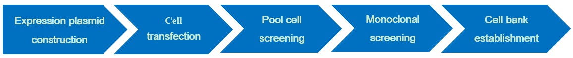
Stable Cell Lines Construction Protocol
Expression plasmid construction
Cell transfection
Pool cell screening
Monoclonal screening
Cell bank establishment
Technical Points
- Expression Plasmid Construction
The base sequence of the target gene with endonuclease site is synthesized by polymerase chain reaction (PCR) method, and then ligated with the vector to construct an expression plasmid. In order to ensure the accuracy of the target gene cloned into the vector, the target gene in the expression plasmid needs to be sequenced before the host cell is transfected.
- Cell Transfection
The expression plasmid is transduced into mammalian cells through four different methods (chemical method, physical method, liposome transfection, viral transfection). The most commonly used methods are electroporation transfection, liposome transfection and poly Ethyleneimine-mediated transfection. The success rate of transfection depends on cell viability, plasmid DNA purity, transfection reagents and transfection method.
- Pool Cell Screening
The cell pools need to be screened and evaluated. The transfected cells should be cultured in a selective medium. When the cells completely restore their viability, they are used to form stable cell pools. The use of reagents depends on the antibiotic resistance gene carried by the expression vector and the type of host cell. For CHO K1 cells, MSX is usually added to the medium without glutamine for selection. While CHO-DG44 cells require glycine, hypoxanthine and thymidine (GHT) for growth. This requirement allows for the selection of recombinant clones by transfection of recombinant DNA plasmids containing a gene of interest and the replacement dihydrofolate reductase (dhfr) gene if cells grow on GHT minus medium then they have taken up the recombinant plasmid DNA and hopefully the gene of interest as well. Increase the selective pressure of methotrexate(MTX) on the DG44 and DXB11 cell pools to obtain the high expression level of target gene. In this screening stage, it is also necessary to evaluate the yield and quality of the target gene expression products, and then select 3-4 cell pools for cloning.
- Monoclonal Cell Colony Screening
The limiting dilution method is mostly used for the traditional monoclonal cell colony screening. It describes a procedure to obtain a monoclonal cell population starting from a polyclonal mass of cells. This is achieved by setting up a series of increasing dilutions of the parent (polyclonal) cell culture. A suspension of the parent cells is made. Appropriate dilutions are then made, depending on cell number in the starting population, as well as the viability and characteristics of the cells being cloned. After the final dilutions are produced, aliquots of the suspension are plated or placed in wells and incubated. If all works correctly, a monoclonal cell colony will be produced.
- Cell Bank Establishment
The establishment of the cell bank is to meet the needs of large-scale production. According to the bioreactor evaluation results and cell line stability research data, select engineered cell lines and candidate cell lines for production. Then establish the primary cell bank PCB, master cell bank and working cell bank under GMP conditions.
Inquiry
