- Home
-
Screening
- Ionic Screening Service
-
Ionic Screening Panel
- Ligand Gated Ion Channels
- Glycine Receptors
- 5-HT Receptors3
- Nicotinic Acetylcholine Receptors
- Ionotropic Glutamate-gated Receptors
- GABAa Receptors
- Cystic Fibrosis Transmembrane Conductance Regulators (CFTR)
- ATP gated P2X Channels
- Voltage-Gated Ion Channels
- Calcium Channels
- Chloride Channels
- Potassium Channels
- Sodium Channels
- ASICs
- TRP Channels
- Other Ion Channels
- Stable Cell Lines
- Cardiology
- Neurology
- Ophthalmology
-
Platform
-
Experiment Systems
- Xenopus Oocyte Screening Model
- Acute Isolated Cardiomyocytes
- Acute Dissociated Neurons
- Primary Cultured Neurons
- Cultured Neuronal Cell Lines
- iPSC-derived Cardiomyocytes/Neurons
- Acute/Cultured Organotypic Brain Slices
- Oxygen Glucose Deprivation Model
- 3D Cell Culture
- iPSC-derived Neurons
- Isolation and culture of neural stem/progenitor cells
- Animal Models
- Techinques
- Resource
- Equipment
-
Experiment Systems
- Order
- Careers
- Home
- Symbol Search
| Catalog | Product Name | Gene Name | Species | Morphology | Price |
|---|---|---|---|---|---|
| ACC-RI0106 | Human KCNMA1/KCNMB1 Stable Cell Line-CHO | KCNMB1 | Human | Epithelial-like | INQUIRY |
Large conductance, voltage, and calcium-sensitive potassium (MaxiK) channels are the basis for the control of smooth muscle tone and neuronal excitability. The MaxiK channel can be formed by two subunits: pore-forming α subunit and Ca2+ /voltage sensing β subunit.
Studies have found that MaxiK channels are sensitive to the external application of several peptidyl toxins, such as charybdotoxin (ChTX), which bind to receptor sites located in the vestibule outside the channel with high affinity and block potassium channels by physically blocking the pores. The researchers found that ChTX specifically binds to the 31-kD β subunit of the MaxiK channel of bovine tracheal smooth muscle. Later, they cloned the cDNA encoding the bovine β subunit and found that the predicted protein contained two putative transmembrane domains. Under non-denaturing conditions, antibodies against the beta subunit immunoprecipitate the alpha and beta subunits of the channel, indicating that the MaxiK channel exists as a polymer containing alpha and beta subunits in vivo. Among them, the KCNMB1 gene encodes a subtype of a beta subunit of the MaxiK channel.
Using the sequence of the bovine β subunit, the scientists cloned the cDNA encoding the human β subunit. Through Northern blot analysis, it was found that human β subunit genes are widely expressed as 1.4-kb transcripts throughout human tissues. And the highest level of expression of this transcript was observed in the aorta. Further analysis revealed that the β subunit genes encoded by mouse Kcnmb1 and human KCNMB1 each contained 4 exons, spanning 9 kb and 11 kb of their respective genomes. And the connection between exons and introns is highly conserved between humans and mice.
The membrane protein family KCNMB has four family members, all of which change the characteristics of MaxiK channels. BK is a widely expressed potassium channel, which can reduce the influx of Ca2+ from voltage-gated Ca2+ channels by hyperpolarizing the cell membrane in response to locally elevated Ca2+. In vascular smooth muscle, BK channels work together with KCNMB1 (β1), which greatly increases their calcium sensitivity. This can strictly control blood vessel tension in response to elevated Ca2+.
KCNMB1 and Disease
KCNMB1 and Hypertension

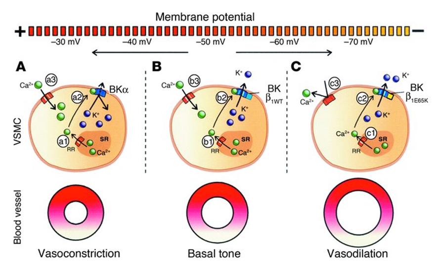
Figure 1. The effect of BK-β1E65K channels in VSMCs. (Fernández-Fernández, José M. , et al.; 2004)
Long-term blood vessels in a state of high blood pressure will cause "ion channel remodeling". The large conductance calcium-activated potassium channel (MaxiK) is the main potassium channel on vascular smooth muscle cells (VSMC). When cell membrane depolarization activates voltage-dependent calcium channels, a large amount of calcium ions influx, and the increase of intracellular calcium ion concentration activates MaxiK channels, generating a large amount of transient outward potassium ion current, cell membrane hyperpolarization, voltage-dependent calcium channel closure, calcium Ion influx decreases and smooth muscle relaxes, so MaxiK channel plays an important role in negative feedback regulation of vascular smooth muscle tension. At present, whether the change of MaxiK channel activity is a cause or a result of hypertension has not yet been unified. Because the MaxiK channel regulates resting potential and vascular tension, many scholars believe that the disorder of MaxiK channel expression causes the occurrence and development of hypertension. Using gene knockout technology, the α and β subunits on the MaxiK channel were used as target genes. The results of the study showed that the deletion of α and β subunits caused an increase in blood pressure in rats. Amberg et al. found that the β1 subunit of the MaxiK channel on the VSMC of spontaneously hypertensive rats (SHR) decreased, resulting in decreased calcium ion sensitivity of the channel and decreased MaxiK channel activity. However, there are also literature data showing that hypertension induces electrical remodeling of VSMC Max iK channels. The cerebral arteries, aorta, carotid arteries and lower limb arteries of SHR all have the increased activity of MaxiK channels.
References
- Harder DR, et al.; Altered membrane elec trical properties of smooth muscle cells from small cerebral arteries of hypertensive rats. Blood Vessels. 1983, 20( 3) : 154-160.
- Sausbier M, et al.; Elevated blood pressure linked to primary hyperaldosteronism and impaired vasodilation in BK channel-deficient mice. Circulation. 2005, 112(1) : 60-68.
- Plüger S, et al.; Mice with dis rupted BK channel beta1 subunit gene feature abnormal Ca(2+) spark/ STOC coupling and elevated blood pressure. Circ Res. 2000, 87(11) : E53-E60.
- Amberg GC, Santana LF. Downregulation of the BK channel beta1 subunit in genetic hypertension.Circ Res. 2003, 93(10) : 965-971.
- Fernández-Fernández, José M. , et al.; Gain-of-function mutation in the KCNMB1 potassium channel subunit is associated with low prevalence of diastolic hypertension. Journal of Clinical Investigation. 2004, 113(7), 1032–1039.
Inquiry
